INTRODUCTION
Intraoperative neurophysiological monitoring (IONM), which refers to the intraoperative use of neurophysiological studies for the functional assessment of neural networks and monitoring of neural integrity during surgery, was introduced in South Korea in the 1990s. However, its use did not become widespread until the 2010s when the Korean National Health Insurance Service (KNHIS) started to cover it, and with the development of a novel modality. In response to the growing popularity of IONM, which is now also a global phenomenon, the Korean Society of Clinical Neurophysiology revised its neurophysiological testing guidelines to include IONM content. The rapidly changing medical landscape both domestically and outside South Korea has necessitated the development of a more practical set of IONM guidelines.
Numerous studies on IONM have been reported, and especially in the official journal of the Korean Society of Intraoperative Neurophysiological Monitoring: the Journal of Intraoperative Neurophysiology. However, these studies do not completely encompass the recent rapid changes in the field of IONM. Accordingly, an IONM quality-management task force was formed to develop the present clinical practice guidelines, which have been tailored to the current IONM conditions in South Korea. These guidelines explain the principles and rationale of IONM together with its supervision, and proposes several recommendations for performing the recently developed diverse IONM techniques.
BASIC PRINCIPLES
The goal of IONM is either the early detection of intraoperative nerve damage or the precise localization of major nerves that are susceptible to intraoperative damage, thereby minimizing the risk of postoperative neurological deficits. Toward that goal, IONM involves monitoring of neural pathways, cerebral blood flow, and the level of neural function during surgery. This not only ensures the safety of patients, but also helps them, their legal guardians, and their medical team to confidently approach the surgery, especially if that surgery is particularly technically challenging.
IONM uses diverse neurophysiological testing techniques that have been developed to diagnose patients with neurological disorders. The types of neurophysiological testing that have been adapted for IONM include motor evoked potentials (MEPs) for monitoring the descending motor pathways, somatosensory evoked potentials (SSEPs) for ascending sensory pathways, brain auditory evoked potentials (BAEPs), visual evoked potentials (VEPs), and electroencephalography (EEG) for recording the electrical activity of the brain, and electromyography (EMG) for monitoring peripheral neuromuscular systems. Transcranial Doppler (TCD), which is a type of ultrasonography, can also be used. Among these diverse modalities, appropriate tests of choice can be chosen according to the particular type of surgery and its inherent risks.
INDICATIONS
In principle, IONM can be used for all surgical procedures for which there is a risk of intraoperative neural damage. However, it is generally only implemented for surgeries with a high risk of postoperative neural deficit or for which evidence-based efficacy of IONM has been demonstrated. These surgeries include functional neurosurgery, such as brain or cerebral nerve surgery, cerebrovascular surgery, epilepsy surgery, and various types of spine and spinal-cord surgery. In general, the involved IONM neurophysiologist will discuss the planned surgery with the surgeon ahead of time to agree upon the most appropriate IONM modalities.
The development and advancements in monitoring devices have expanded the target surgical procedures for which IONM can be implemented. At each institution, the IONM modalities and their settings should be decided for the target surgical procedure at the discretion of the neurophysiologist and with the cooperation of the anesthesiologist and surgeon.
THE IONM TEAM
It is essential that the IONM team includes neurophysiologists, operating surgeons, anesthesiologists, and medical technologists. A close interaction between these members is required for precise and successful IONM.1
The neurophysiologist is responsible for the overall management of IONM, including the choice of IONM equipment, settings, and monitoring method for each case, and for determining the level of monitoring that will be required, interpreting IONM results, and educating the monitoring technologist.2 As a supervising physician, the neurophysiologist should be familiar with a wide range of monitoring techniques, know which is the most appropriate to obtain the required waveforms during a particular type of surgery, recognize the meaning behind any changes in those waveforms and be able to reduce any artifacts, and be able to supervise and guide the medical technologist in the performance of these monitoring techniques. In this setting, neurophysiologists should also be able to not only properly and clearly report waveforms or waveform changes observed on a monitor to the operating surgeon, but should also be able to interpret their clinical significance. They should not only have a good understanding of neurophysiology, but should also have received full-time training on the methods of electrophysiological testing including generation of evoked potentials (EPs), EMG, EEG, nerve conduction studies (NCS), and ultrasonography. In South Korea, board-certified neurologists or rehabilitation medicine physicians would qualify as neurophysiologists based on the training that they receive.
A medical technologist is defined by Korean domestic laws as a professional who is supervised by a physician or a dentist and is responsible for performing chemical or physiological tests. As a member of an IONM team, the medical technologist performs neurophysiological tests in the operating room. IONM does not refer to a specific type of test, but rather to the comprehensive use of a collection of various clinical neurophysiological tests such as generating EPs, EMG, EEG, NCS, and TCD. Simply knowing how to use IONM equipment or having the ability to interpret IONM waveforms does not necessarily indicate that the neurophysiologist has received sufficient training to perform optimal IONM.
While a full IONM team consisting of at least four members- an operating surgeon, an anesthesiologist, a neurophysiologist, and a medical technologist trained by a neurophysiologist-is usually necessary to perform IONM, a smaller team may suffice under limited circumstances. For instance, a single operating surgeon with a single anesthesiologist may be sufficient for thyroid surgery, whereby the operating surgeon locates the recurrent laryngeal nerve using an automated device, performs vagus nerve stimulation, and uses a device that records vocal-cord movements from recording electrodes and converts the generated EMG signals into sounds. However, the IONM process as performed by a small number of medical professionals should differ from that performed by a complete team in terms of its purpose and accuracy, and therefore should not be considered equivalent. Furthermore, alarm sounds or simple verbal communication about any waveform changes are not recommended for interpreting waveforms in IONM if they are not accompanied by the interpretation of the waveforms observed on a monitor. A neurophysiologist who can perform and supervise IONM and a medical technologist certified in neurophysiology are essential members of an IONM team. It is recommended that these personnel cooperate with the operating surgeon and anesthesiologist as part of an integrated IONM team.
LEVELS OF SUPERVISION AND INSURANCE BILLING
Defining the required level of supervision by a clinical neurophysiologist during IONM is important, and is summarized as below based on recommendations from medical communities in foreign countries and textbooks on IONM.
Supervision of medical technologists by a neurophysiologist during IONM is classified as direct, personal, or general depending on the purpose and method of supervision. Among these, direct supervision is the most commonly used during IONM according to current KNHIS requirements. In performing direct supervision, a neurophysiologist should be available close to the operating surgeon or medical technologist to provide supervision when needed, or should provide online supervision in real time. The potential for direct online supervision is currently increasing due to recent advances in communication technology and devices. In South Korea, direct online supervision is provided during IONM for most surgical procedures involving the brain, spine, spinal cord, and blood vessels. In general, routine tests performed in a laboratory (e.g., MEPs and SSEPs) are repeated to monitor waveform changes, and the test results are objectively represented as numbers or waveforms changes. Most textbooks as well as domestic and foreign recommendations state that IONM does not require a neurophysiologist to remain in the operating room throughout the surgery. For example, the coverage policies recently revised by the American Academy of Neurology state that supervision is performed either in the operating room or via a real-time connection outside the operating room.3 Therefore, it is sufficient for a neurophysiologist to perform real-time monitoring close to the operating room (usually within the same building where the surgery is taking place or somewhere nearby) via a wired (local area network or optical cable) or wireless connection, and provide immediate supervision to a medical technician when major changes in waveforms occur The neurophysiologist is responsible for interpreting the waveform changes, and enters the operating room only when necessary.4-8
Personal supervision is provided for IONM that includes brain stimulation and cognitive tests such as assessment of the language and motor regions of the brain for brain tumor or epilepsy surgery. These require high levels of professional knowledge and experience. It is recommended that a medical technologist performs IONM under the personal supervision of a neurophysiologist.4-6 However, extensive discussions on the testing method and interpretation of the results, as well as communication between the operating surgeon and the neurophysiologist are necessary before performing IONM that requires personal supervision. The medical cost for personal supervision during IONM is not currently specified by the KNHIS. General supervision is provided by a neurophysiologist in the absence of direct or personal supervision of a neurophysiologist.
While IONM is performed simultaneously and in the same space as the surgery and anesthesia, it remains distinct from both. Therefore, in countries including the United States, neither the surgeon nor the anesthesiologist performing a surgical procedure can bill for IONM since it is included in the global package when serving as the physician supervising IONM.3 It is not advisable for a neurophysiologist to provide direct supervision for IONM performed on an outpatient basis. Given that neurophysiologists in South Korea are also responsible for providing outpatient services in departments of neurology or rehabilitation medicine, it is recommended that each institution has at least two neurophysiologists to allow IONM to be performed continuously. It is also recommended that neurophysiologists maintain appropriate levels of supervision with respect to both the patient and the surgical procedure during IONM.
PRECAUTIONS
Safety
In IONM, electrical stimulation is commonly performed using rectangular pulses. The amount of electrical charge delivered (Q, mC) is calculated by multiplying the equivalent pulse current (I, mA) by the duration of the pulse (D, msec): Q = I × D. The delivered electrical charge is the most important stimulation measure related to neurological damage. The amount of energy (E, mJ) delivered is related to the total resistance (R, kOhm) and is calculated as E = I2 × D × R × 0.0001. For safe electrical stimulation, the intensity of the current is generally inversely proportional to the duration of stimulation, which is generally set to between 0.02 and 1 msec. The current threshold decreases dramatically as the duration of the stimulating pulse decreases, and the threshold charge and energy also decrease in proportion to the duration. Therefore, it is important to set the duration and current below their threshold values when performing electrical stimulation. While the electrical stimulation devices that are currently available on the market prevent the user from applying excessive stimuli for safety reasons, the user should still use appropriate stimulation settings after considering the condition of an individual patient.
For MEPs, transcranial electric motor evoked potentials (TCE-MEPs) are commonly measured; these are generated by applying a strong electrical stimulus to the cerebrum through the cranial bone. Transcranial electrical stimulation causes seizures (rarely, 1 in 3,000 patients), or tongue or jaw injuries due to electrical-stimulation-induced oral muscle contractions. Having a pacemaker or any other device inside the body is also a potential contraindication for transcranial electrical stimulation.9 When delivered at higher intensities, these stimuli can reach the deeper brain structures.10 Therefore, the lowest stimulus intensity should be considered, or else MEPs generated by direct cortical stimulation should be examined in surgeries where it is necessary to detect small-nerve damage in the cerebral cortex.
For direct cortical stimulation performed to assess cortical function, continuous single-pulse stimulation may be used (Penfield method), whereby an electrical stimulus consisting of a single pulse is given for < 10 seconds; alternatively, stimulation with a train of multiple pulses may be considered. SSEPs are generated by electrically stimulating peripheral nerves, and can be safely performed in the outpatient clinic. It is recommended that the appropriate IONM tests, stimulation methods, and stimulation thresholds should be chosen separately for each individual patient.
Effect of anesthesia
Anesthetic agents can significantly affect waveform generation during IONM. Total intravenous anesthesia (TIVA) using propofol and low-dose opioids is the most widely used anesthetic technique for surgeries in which IONM is planned. TIVA is reported to be superior to existing anesthetic techniques in terms of MEP generation and waveform size in IONM. However, even with TIVA, an excessively high dose of an intravenous anesthetic agent can reduce MEP generation and the size of EEG waveforms.11 Ketamine or low-dose etomidate can increase the size of SSEP or MEP waveforms, although these are no longer commonly used.12
Neuromuscular relaxants are commonly used during surgery to facilitate the insertion of an endotracheal tube and to reduce muscle tone. Since muscle relaxants can result in partial or complete loss of MEP or EMG signals in IONM, it is recommended that a short-acting neuromuscular relaxant be used (e.g., rocuronium) just once, immediately before tube insertion. Stimulation with a train of four pulses can be used to easily detect any remnant neuromuscular relaxants in the body.1
Given the impact of anesthesia on waveform generation, it is strongly recommended that the effects of different anesthetic agents on IONM be discussed with the anesthesiologist prior to the surgery so that the most appropriate can be chosen according to the type of IONM tests that are planned.
IONM with optimal waveform generation is performed while maintaining the level of anesthesia at which the surgery can be safely performed. Caution is recommended when using muscle relaxants while performing IONM for motor pathways, and also when using anesthetic inhalants while performing IONM for cortically generated waveforms.
ELECTRODES AND EQUIPMENT
IONM device
While well-established neurophysiological testing techniques that have been used for outpatients clinics are adapted for IONM, those techniques can be significantly affected by anesthetic agents, the use of different devices, movements of the operating surgeon, and the absence of electrical shielding in the operating room. Therefore, equipment specifically designed for IONM is generally recommended over that used to examine outpatients or inpatients outside of the operating-room environment. Various IONM devices are available domestically and outside of South Korea, and it is recommended that the most appropriate device be chosen according to the needs and characteristics of individual centers.
Electrodes
Various types of electrode can be used for IONM. Needle electrodes are the most common, as they are convenient to use and because patients are under anesthesia when the electrode is placed. Needle electrodes can be substituted by cup or surface electrodes, which are used for obtaining EEG signals and SSEPs in outpatients. Once detached, needle electrodes disrupt waveforms in IONM and could injure or cause infection in the personnel performing the procedures. It is therefore recommended that a bandage, plaster, or tape be placed over the needle-insertion site following placement of the electrode. Extra caution is required to prevent infection in the patient when fixing the electrodes near to the surgical site.
Other types of electrode can be used for more-active IONM. Surface electrodes that are attachable or fixed to the endotracheal tube can record EMG signals from the vocal-cord muscles, which cannot be easily recorded by common surface or needle electrodes. They may be useful for the early detection and prevention of damage to the vagus nerve and recurrent laryngeal nerve following surgical procedures to the lower brainstem or thyroid or parathyroid glands.13 Hook-wire electrodes can be safely used to record EMG signals from the small muscles for which the installation of subcutaneous electrodes could be dangerous. Specially designed electrodes may be used to record compound nerve action potentials from the sphincter muscle of the anus or from cranial nerve endings. Subdural electrodes or epidural electrodes can be used to record electrical potentials through direct contact with the cerebral cortex and spine.
METHODS
Preparation
a) The IONM team should be aware of the patient’s clinical details, including their diagnosis, type of surgery to be performed, and preoperative neurological condition.
b) The clinical neurophysiologist should discuss the required IONM modalities and the most efficient and most appropriate anesthetic technique with the operating surgeon and anesthesiologist. A consensus regarding these may be reached prior to surgery or formalized for a particular diagnosis/surgical procedure, and should be clearly delivered to the clinical neurophysiologist.
c) After definitively identifying the patient, the IONM team should record this information on the IONM record before anesthesia induction is complete.
d) Once the patient is fully anesthetized, electrodes are placed and fixed in place. After adjusting the patient’s position for the surgery, the electrodes are loosened and connected to the IONM device.
e) After all of the electrodes are connected to the device, an impedance test is performed and baseline waveforms are recorded. These waveforms provide information about the depth of anesthesia and whether the IONM device is operating properly.
Attaching electrodes and sensors
a) Transcranial electric stimulation is usually performed using needle electrodes and, rarely, using those with a corkscrew design. Electrodes can be placed at Cz, C3/C4, or C1/C2 depending on the location of the muscle being stimulated. It has been suggested that at relatively low stimulation intensities, placing electrodes closer to the medial side (i.e., Cz) facilitates the recording of MEPs from the leg muscles, whereas placing them closer to the lateral side (i.e., C3/C4) facilitates the recording of MEPs from the arm muscles.14 However, caution is required since placing electrodes too close to the lateral side can lead to tongue or jaw injuries due to contraction of the oral muscles.
b) The procedure for electrical stimulation for inducing SSEPs, BAEPs, VEPs, and EMG signals is the same as that used during the diagnostic examination of patients with neurological conditions.
c) EEG and TCD are performed using a procedure similar to that used in the outpatient clinic. Scalp electrodes for EEG should be placed according to the international 10-20 system. Subdural electrodes are used to record the electrocorticography (ECoG). For TCD, the head band for the probe is placed at a location where the blood flow in the middle cerebral artery can be measured using a 2-MHz probe.
Stimulation
a) To measure TCE-MEPs, short trains of five to seven electrical pulses are transmitted through the electrodes placed on the scalp. This continuous electrical stimulation results in the firing threshold of alpha motor neurons being reached even at low stimulus intensities, and can generate MEPs in patients under appropriate levels of anesthesia.15
b) The stimulation procedure for SSEPs, BAEPs, and VEPs is similar to that used in the outpatient clinic. However, in the case of SSEPs, subdural electrodes can also be placed on the wrists and ankles near to the nerves to be stimulated instead of placing the stick-type electrodes used in the outpatient clinic. For BAEPs, tubal insert phones are inserted to stimulate nerves with the aid of a clicking sound. For VEPs, flashlights or LED lights that do not require a hairband for fixation are easy to install, and generate a small and bright stimulus that can be used during surgery.
c) Contrary to the tests performed in the outpatient clinic, electrical stimuli are transmitted to the cranial nerves or nerve roots after exposing the peripheral nerves in the operating room during the stimulation of peripheral nerves to record compound motor action potentials from the muscles.
d) Electrical stimuli can be applied locally via bipolar stimulation or over a wide area via unipolar stimulation.
e) It is important to tailor the type and method of stimulation for each patient to the type of surgery and the monitoring technique being used.
Recording
Baseline data
In neurophysiological tests performed for diagnostic purposes, measurements are compared with normal values to detect any abnormalities while taking into account the patient’s general characteristics such as age or height. In IONM, baseline waveforms measured before the main surgical procedure are compared with those obtained during surgery to detect any surgery-related changes in neurological function.
Since IONM waveforms are significantly affected by anesthetic agents, the patient’s general condition, and proper functioning of the IONM equipment, it is advisable to obtain baseline IONM waveforms when 1) the patient has been anesthetized; 2) the injection rate of a TIVA intravenous agent becomes constant; 3) there is no effect of neuromuscular blockers; and 4) the patient does not have severe bleeding, hypothermia, or electrolyte imbalance. It is also advisable to obtain several baseline waveform measurements and choose the most reproducible results among those waveforms for comparison with those obtained during intraoperative recording.
TCE-MEPs
Transcranial electric muscle motor evoked potentials (TCE-mMEPs) are usually recorded from the muscles. While the electrode installation for TCE-mMEP measurements is simple, there can be high intertrial variabilities between each TCE-mMEP recording.16 The D-wave technique was developed to overcome these variabilities, whereby MEPs are recorded directly from the spinal cord, with the claim of more-accurate detection of intraoperative nerve damage compared with TCE-mMEPs. However, since studies on the efficacy of the D-wave technique have included relatively small numbers of patients, large-scale prospective research is needed to allow clearer conclusions to be drawn about its advantages over TCE-mMEPs.17 Catheter electrodes specially designed to measure MEPs from the spinal cord in the epidural or subdural space are necessary to measure D-waves.
INTERPRETING AND DOCUMENTING RESULTS
When an alarm sound alerts to the possibility of nerve damage during surgery, the clinical neurophysiologist and medical technologist should quickly investigate whether the IONM device is functioning properly, check for artifacts, and check for changes in the anesthetic agents or the patient’s general condition (blood pressure, body temperature, oxygen saturation level, electrolyte levels). If none of these are observed, they should inform the operating surgeon of the possibility of intraoperative nerve damage.
Criteria for alarms
When examining blood flow changes using waveforms, the following are checked in the given order: a reduction in the velocity of the fast wave, increased slow waves, reduced amplitude, burst suppression, and even flat EEG signals in severe cases. Blood flow is considered abnormal if there is a ≥ 50% reduction in the amplitude of the slow wave. ECoG may be performed to check for the presence of epileptic discharges and locate the site of any blood flow disruption.
TCE-mMEPs
TCE-mMEPs show high intertrial variability between each stimulation, and the optimal alarm criteria to alert to changes in intraoperative TCE-mMEPs are still a matter of debate. The absence of MEPs is reported to predict the possibility of nerve damage in the spine during spinal-cord surgery with the highest specificity. However, partial loss of MEPs or a reduction in the size of MEP waveforms (50%, 70%, or 80%) can still indicate mild postoperative motor deficits in a small number of patients.18
In IONM for brain surgery, a reduction in MEP amplitude often indicates postoperative motor deficits in the absence of the complete loss of waveforms. It has been suggested that a ≥ 50% reduction in the amplitude of an MEP waveform can indicate the risk of intraoperative nerve damage during brain surgery.19
The predictive values of intraoperative MEP changes can depend on the muscles from which the MEPs were measured. For instance, MEPs from the muscles controlled by a large number of corticospinal fibers (e.g., the abductor halluces in the foot) have high specificity, whereas MEPs measured from those controlled by a small number of corticospinal fibers (e.g., tibialis anterior in the leg) have high sensitivity.20
Given that none of the alarm criteria for intraoperative MEPs have 100% specificity and sensitivity, it is necessary to establish alarm criteria appropriate for each IONM center based on the results of previous studies. However, it is noteworthy that alarm criteria bearing high sensitivity are bound to reduce the test’s specificity, and vice versa.
SSEPs
In general, the risk of intraoperative nerve damage is deemed high if the SSEP latency is extended by ≥ 10% or if its amplitude is reduced by ≥ 50%.4
BAEPs
In general, the risk of intraoperative nerve damage is deemed high when V-wave latency is extended by ≥ 1 msec or if its amplitude is reduced by ≥ 50%. It has been suggested that only the complete loss of the wave V indicates a high risk of intraoperative central nervous system damage during, surgery with the exception of surgery for cerebellopontine angle tumors;21 however, further research is needed to confirm this finding.
VEPs
In general, the risk of intraoperative nerve damage is deemed high when there is a ≥ 50% reduction in the amplitude of the P100 wave, a component of the VEP that peaks at 100 msec. Electroretinography may be performed to examine the presence of the P100 wave, which is useful for examining prechiasmatic lesions; its usefulness for assessing postchiasmatic lesions is currently under investigation.
EMG
Neurotonic discharges are the clinically critical EMG signals generated during the cranial nerve and neuromuscular stimulation. These neurotonic EMG signals are more continuous than phasic discharges. They are generated by the repeated firing of motor units and have a high frequency of 50-200 Hz. Since phasic discharges can be produced upon minor stimulation such as touching a nerve or spraying water, they are not considered clinically significant. On the other hand, neurotonic discharges can indicate mechanical, thermal, or metabolic nerve stimulation or damage; however, they should be interpreted with caution as this is not all neurotonic EMG signals indicate intraoperative nerve damage. For instance, Gunnarsson et al.22 studied the association between IONM changes after spinal surgery and postoperative nerve damage and reported that neurotonic EMG signals have 100% sensitivity but only 23.7% specificity in predicting postoperative paralysis after spinal surgery. Electromyographic responses can be roughly examined by listening to the sounds of EMG signals. Repeated, high-pitched sounds are generated in the presence of strong neurotonic discharges. However, this sound-based method is generally not recommended since, as explained earlier, it provides only limited information about the IONM waveforms.
TESTING TIME
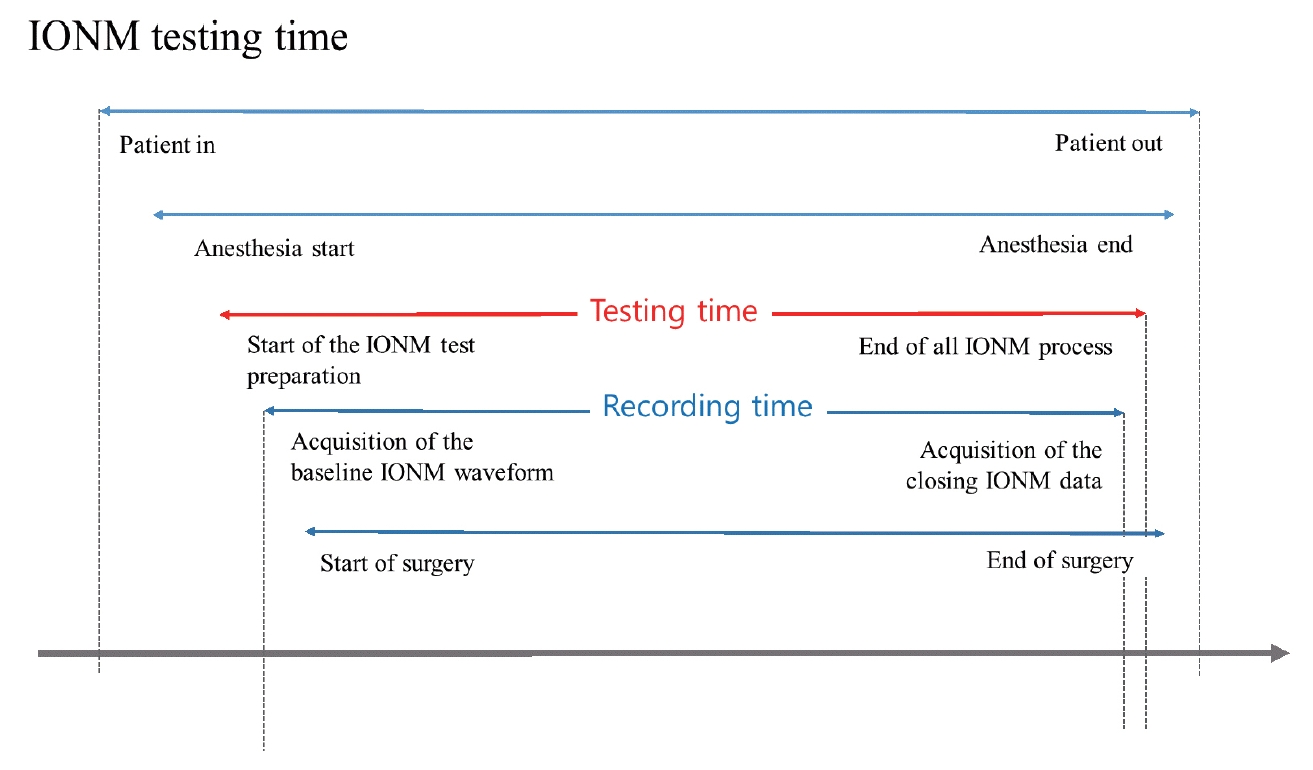
TCD may be used to check for any disruption in the cerebral blood flow based on a reduction in systolic blood flow. High-intensity transient signals generated by emboli as well as high-pitched sound signals may also be used to assess cerebral blood flow.The billing for IONM depends upon the duration of testing, and therefore precise recording of the testing time is important. Both the recording time and testing time are usually referred to in the billing process for IONM (Fig. 1). The recording time refers to the time from the start of recording of baseline waveforms to when the closing data are obtained. The testing time refers to the time from the start of test preparation, including setting up the equipment and placing the electrodes and stimulator, to the time when the IONM process, including electrode removal/disposal, patient examination, and saving of data, is complete. Billing IONM based on the test time is most appropriate since IONM starts before and continues after data collection.
INTERPRETATION
Documentation of data interpretation should show the basic information, the modality of monitoring, the monitoring results, and the conclusions, and may include additional comments if needed. The basic information includes the date of the test, and the patient’s name, sex, age, and registration number. It should also include details about the surgery itself, including the type of surgery, the surgical site, and the name of the operating surgeon, as well as information about the anesthetic procedure, including the type of anesthetic procedure, doses of anesthetic agents, and the names of the anesthesiologist, medical technologist, and neurophysiologist. It is recommended that the monitoring and recording times be indicated clearly when documenting the basic information, in order to facilitate the insurance claims process (Fig. 2).
SUMMARY
These guidelines include useful recommendations that are supported by currently available clinical evidence. They provide explanations of the basic principles, indications, team composition, levels of supervision, precautions, and equipment, including electrodes and other devices for IONM. Moreover, a summary of current monitoring techniques, methods for interpreting and documenting IONM results and recording the testing time, and principles of IONM interpretation are described. Finally, these guidelines provide an in-depth summary of the different levels of supervision, testing times, and methods of IONM data interpretation in relation to KNHIS coverage.