Germ-cell tumors (GCTs) in the spinal cord are exceedingly rare, with only about 44 cases having been reported worldwide.1 They usually present as intramedullary lesions in the thoracic or thoracolumbar spine. Since imaging produces nonspecific findings, diagnosing GCTs is mostly dependent on a pathological confirmation. We present a patient with a primary intramedullary GCT, who was diagnosed after many years and finally underwent radiotherapy and chemotherapy.
CASE
A 22-year-old female presented with gradually progressive weakness of both legs that had first appeared 5 years previously. Her first symptom was her shoes keeping coming off at an age of 17 years. She did not have any voiding difficulty or constipation. The symptoms worsened after 6 months, when she needed to place her hands on the ground in order to stand up. At that time she also had spasticity in both legs. Various tests including a neurological examination and spinal magnetic resonance imaging (MRI) performed in December 2014 revealed no abnormality. Although there was no family history of neuromuscular disorder and gene testing covering Spastin and Atlastin GTPase 1 did not reveal any abnormality, hereditary spastic paraplegia (HSP) was suspected because of its diverse genetic variants and the presence of progressive weakness and spasticity in both legs. The patient was told that rehabilitation was the only suitable treatment. The weakness in both legs continued worsening until she ended up in bedridden state. After 5 years of the illness she visited our hospital for a second opinion.
A neurological examination revealed weakness in both legs symmetrically, which were about Medical Research Council (MRC) Grade III, and MRC grade I distally. She had hypesthesia in both legs to all modalities. Her deep tendon reflexes were reduced in both legs, and spasticity was not definite despite initial spasticity being recorded in her medical records. She also had voiding difficulty and constipation. Cerebrospinal fluid (CSF) tapping showed mild lymphocytic pleocytosis (red blood cell, 2 cells/mm3; white blood cell, 10 cells/mm3) with a normal protein level (0.34 g/L). Atypical cells were not observed in the CSF. Testing for the following HSP genes did not reveal any pathogenic variant: ATL1 (SPG3A), BSCL2, CYP7B1, HSPD1, KIAA0196, KIF5A, L1CAM, NIPA1, PLP1, REEP1, SLC16A2, SPAST(SPG4), SPG11, SPG20, SPG21, SPG7, ZFYVE26 (SPG15), and ZFYVE27.
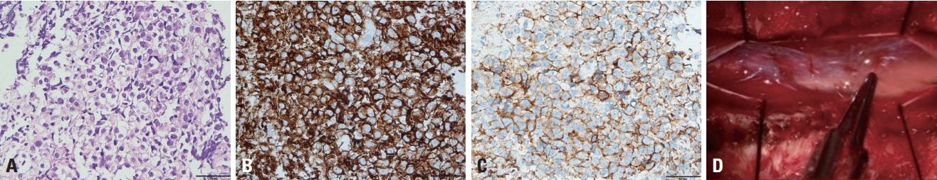
Since the patient’s laboratory and clinical findings were not specific to HSP, a repeat MRI study with contrast medium was performed despite the initial MRI showing no abnormality. Brain MRI showed no structural abnormalities. However, spinal MRI performed in August 2018 revealed a newly developed fusiform swelling at levels T12-L1 of the spinal cord with minimal homogeneous enhancement (Fig. 1A-C). The radiological findings led to astrocytoma as the suspected primary diagnosis. Laminectomy and tumor removal were performed. A grayish purple mass was observed during the surgery, with no definite dissection plane evident between the spinal cord and the mass (Fig. 1D, E).
The tumor was analyzed using hematoxylin-eosin staining, immunohistochemistry, and next-generation sequencing (NGS). The tumor showed positivity for placental alkaline phosphatase and CD117, and 95% positivity for Ki-67. NGS revealed loss of the KIT gene, with 25% of the germinoma being altered. Primary intramedullary spinal cord germinoma was confirmed as the final diagnosis (Fig. 2).
The patient underwent chemotherapy with a combination of etoposide, ifosfamide, mesna, and cisplatin. Radiotherapy was applied at 36 Gy in 20 fractions, and follow-up spinal MRI revealed shrinkage of the tumor. However, significant neurological deficits remained.
DISCUSSION
Primary intramedullary germinoma is rare and usually involves the lower thoracic part of the spinal cord, as in the present patient.2 A GCT looks similar to an astrocytoma in MRI, which can be hypointense or isointense in T1-weighted images, hyperintense in T2-weighted images, and moderately enhanced in gadolinium-enhanced images. The initial imaging typically reveals a mass,2 but in rare cases it can only show spinal cord atrophy, which is an early sign of intramedullary germinoma.3 These features make a radiological diagnosis difficult, and hence biopsy or resection with pathohistological examination is necessary.4
HSP is often misdiagnosed since it may present with various symptoms and the presence of gene mutation might not be verified. Therefore, clinicians must check for other treatable diseases before diagnosing HSP. An intracranial germinoma is a radiosensitive tumor whose cure rate is more than 90% with radiotherapy alone,5 which is also the case for intramedullary germinoma.2 The response to treatment is highly favorable, but an early diagnosis of germinoma is critical for a good prognosis. It is particularly important to note that early imaging findings might not be apparent, as was the case in the present patient. Thus, if the diagnosis is not clear, MRI follow-up is necessary. Although rare, germinoma should be considered in the differential diagnosis of young adult patients with a tumor in the thoracic or thoracolumbar spine.